Hepatic portal vein labeled 209633-Hepatic portal vein labeled
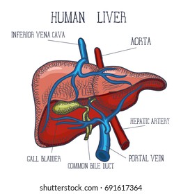
The portal vein is formed by the union of the superior mesenteric vein and the splenic vein just posterior to the head of the pancreas at about the level of the second lumbar vertebra It extends slightly to the right of the midline for a distance of 55–8 cm to the porta hepatis The portal vein has a segmental intrahepatic distribution, accompanying the hepatic arteryThere's a blood vessel that carries blood from the gastrointestinal tract to the liver Here's the story of that blood vesselDaily Anatomy AppFor a random Portal vein lined by endothelium Bile duct lined by cuboidal epithelium, approximately the same diameter as a hepatic artery Not every normal portal tract will always show an artery, vein and bile duct;

Left Gastric Vein An Overview Sciencedirect Topics
Hepatic portal vein labeled
Hepatic portal vein labeled-This is the preview of our full video about the hepatic portal vein Learn more about one of the most important vessels in the human body and watch our fullB – Vein C – Capillarity ii The parts labelled 1 to 3 are 1 Connective tissue layer 2 Lumen 3 Muscular layer iii Name the type of blood that flows through A Oxygenated blood iv The difference is A is an artery and has thick muscular walls which are elastic in nature B is a vein which has thin muscular walls which are less elastic in nature v
:max_bytes(150000):strip_icc()/GettyImages-188057933-5999a71d685fbe0010fa2663.jpg)


Portal Vein Anatomy Function And Significance
Name the veins labelled 19 Trace the flow of blood through them, including the meetup points of veins 1 Hepatic vein 2 Hepatic sinus 3 Hepatic portal vein 4 Gastric vein 5After passing through the liver, the blood collects and leaves in hepatic veins These major blood vessels, enter and leave the liver at the porta hepatis Also emerging from the porta hepatis are the left and right hepatic ducts which contain the collected bile, and the efferent lymphaticsThe hepatic portal vein is formed by the confluence of three main vessels, the gastric, pancreaticomesenteric, and lienomesenteric veins They unite to form the hepatic portal vein near the anterior tip of the dorsal lobe of the pancreas Recall that the celiac artery splits into its branches very near this point as well
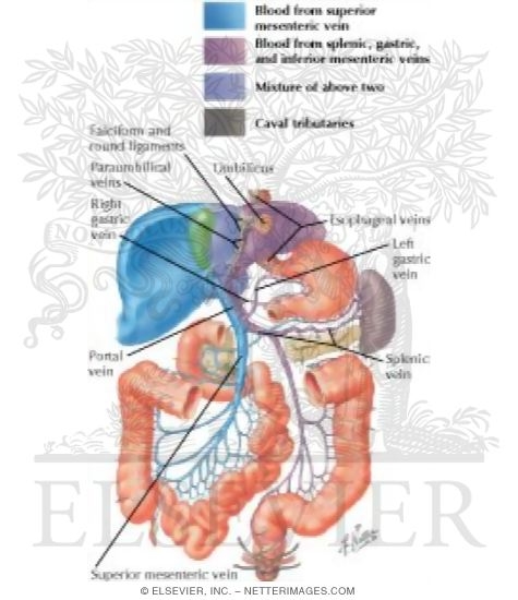
The hepatic portal vein is the largest vein in the abdominal cavity It drains blood from the spleen and the gastrointestinal tract to the liver The hepatic veins begin at the junction of splenic veins and superior mesenteric The blood from the cystic veins and the inferior mesenteric gastric veins is also drained by the hepatic veinHepatic vein, posteriorly bounded by the plane of the right hepatic left portal veins, with the right portal vein subsequently dividing into anterior and posterior branches Standard anatomy (6580%) Main portal vein trifurcation into right anterior, rightThrombosis of the portal vein MED die Pfortaderthrombose pressure in the portal vein MED der Pfortaderdruck stenosis of hepatic veins MED Block der venösen Leberstrombahn portal die Portalseite Pl die Portalseiten portal die Pforte Pl die Pforten portal der Portalrahmen Pl die Portalrahmen vein kein Plural style, tendency der
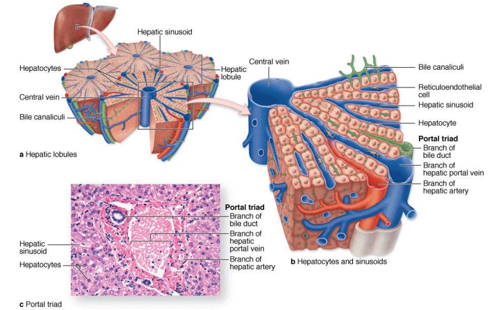
Right hepatic vein The longest of the hepatic veins, the right hepatic vein and lies in the right portal fissure, which divides the liver into an anterior (frontfacing) and posterior (rearfacing) sections Middle hepatic vein This vein runs at the middle portal fissure, dividing the liver into right and left lobes Hepatic portal vein (histological slide) Together with the bile duct, the hepatic portal vein and hepatic artery form the portal triad Those structures supply blood to the sinusoids and the hepatocytes, subsequently draining into the central vein followed by the sublobular veinsLinked Keywords Images for Liver Model Labeled Hepatic Portal Vein LIVER ANATOMY Diagnostic Medical Sonography Abdominal amazonawscom Untitled Document biosunyorangeedu sunyorangeedu


The Hepatic Portal System Flashcards Quizlet


1
The portal vein (PV) is the main vessel of the portal venous system (PVS), which drains the blood from the gastrointestinal tract, gallbladder, pancreas, and spleen to the liver There are several variants affecting the PV, and quite a number of congenital and acquired pathologies In this pictorial review, we assess the embryological development and normal anatomy of the PVS,View Labeled Veins on Bloody Billdocx from ANAT 1572 at College of DuPage Subclavian Vein Internal Jugular Vein (L) Superior Vena Cava Brachiocephalic Vein Hepatic Portal Vein Axillary Vein Hepatic segmentation We outlined the various hepatic segments by partitioning the segment IV into IVa and IVb We defined the segments using the subhepatic veins Human Anatomy display of anatomical labels To use this module, you must click on the "anatomical structures" tab You can then choose between the different structures available


Hepatic Veins Wikipedia


1
The portal fraction (PF) of total hepatic blood flow was calculated in all patients using indicator dilution curves obtained from the portal bifurcation, a right hepatic vein, and when possible a left hepatic vein (six cases) after injection of 51 Crlabeled red blood cells (51 Cr RBC) into the superior mesenteric arteryID Title GI Anatomy Portal System Category LabeledMulroney Flash Cards ID 5058 Title Hepatic Portal vein Tribu Category LabeledMultiple PublicationsOrigin Hepatic Portal Vein is formed by the union of Splenic vein and Superior mesenteric Vein behind the neck of pancreas at L1 vertebral level Termination The portal vein terminates by branching into right branch (entering right lobe of liver) and left branch entering (left lobe of liver) Tributaries Splenic vein;
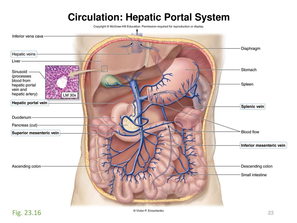


Activity 10 Vessels And Circulation Ppt Download
:background_color(FFFFFF):format(jpeg)/images/library/13688/dZNM4CXK8lO7NsLAptEGw_Left_gastric_vein_2.png)


Left Gastric Vein Anatomy Tributaries Drainage Kenhub
The hepatic portal vein is one of the most important vein that receives blood from the body and transports it into the liver for filtration and processing This vein is part of the hepatic portal system that receives all of the blood draining from the abdominal digestive tract, as well as from the pancreas , gallbladder , and spleenIt supplies approximately 75% of the liver's blood The hepatic arteries supply arterial blood to the liver and account for the remainder of its blood flowCentral veins are quite prominent and provide an easy means of orientation in sections of liver At the vertices of the lobule are regularly distributed portal triads (also known as portal tracts) Examination of a triad in cross section should reveal a bile duct and branches of the hepatic artery and hepatic portal vein


Flat Wire Model
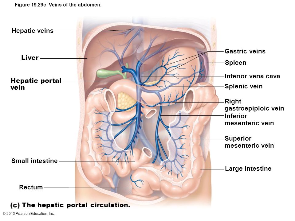


33 Label The Veins Of The Hepatic Portal System Labels Design Ideas
Hepatic veins Transverse diagram of the liver shows the right hepatic vein (RHV), middle hepatic vein (MHV), and left hepatic vein (LHV) draining into the retrohepatic inferior vena cava (IVC) The hepatic veins divide the liver into Couinaud system segments as indicated The hepatic veins are interro The hepatic portal system is the system of veins comprising the hepatic portal vein and its tributaries The liver consumes about % of total body oxygen when at rest, so the total liver blood flow is quite high Blood flow to the liver is unique in that it receives both oxygenated and partially deoxygenated bloodThe hepatic portal vein carries venous blood drained from the spleen, gastrointestinal tract and its associated organs;
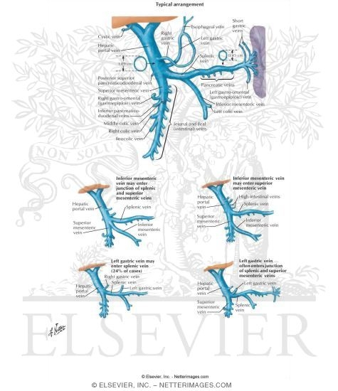


Variations Of Hepatic Portal Vein


Http Www Cccprofessorlou Com Resources Biology 2341 Lab Materials Diagrams arteries and veins Pdf
Portal Triad Anatomy Made up of the lesser omentum that surrounds the common bile duct (CBD), hepatic artery, and portal vein It extends down between the lesser curvature of the stomach and the groove where the ligamentum venosum residesAnat portal vein Vena portae Pfortader {f} anat hepatic vein Vena hepatica Lebervene {f} med occlusion of the portal vein Pfortaderverschluss {m}LhvLeft hepatic vein ivc Inferior vena cava Parasagittal Midclavicular RPV Right Portal Vein RHV Right hepatic vein Parasagittal Right Porta hepatis is seen with an oblique angle 45degree rotation from the sagittal view to the transverse view Oblique left showing the ligamentum teres Transverse Plane showing the Ligamentum Venosum
:max_bytes(150000):strip_icc()/GettyImages-188057933-5999a71d685fbe0010fa2663.jpg)


Portal Vein Anatomy Function And Significance
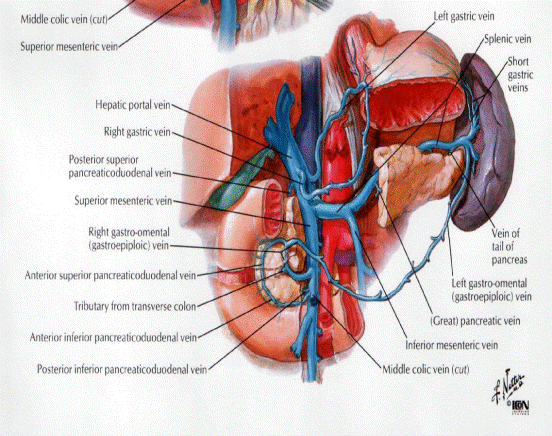


What Vein Drains The Lesser Curvature Of The Stomach And Empties Directly Into The Hepatic Portal Vein Socratic
An ultrasound can be used to detect some liver conditions The function of the hepatic portal system is to carry blood into the liver through the hepatic portal vein from small blood vessels called capillaries located in parts of the abdomen This vein carries blood from the intestines, pancreas, stomach, and spleen to sinusoids, or dilated capillaries, in the liverLiver Model Labeled Hepatic Portal Vein Loading AZ Keywords Keyword Suggestions liveresult;Some will show multiple artery or duct faces (Hepatology 1998;223) Lymphatics not normally seen on H&E


Untitled Document
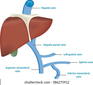


Hepatic Portal Vein High Res Stock Images Shutterstock
ID Title GI Anatomy Portal System Category LabeledMulroney Flash Cards ID Title Hepatic Portal Vein Tribu Category LabeledTrelease Surgical AnatomyBody Quadrants Labeled hepatic portal vein anatomy Sonography ️ When using this image in external sources it can be cited as these quadrants are defined by where the umbilical plane and the sagittal plane intersect But there are also plotted labeled ordered pair of numbers that go on the quadrants The hepatic portal vein carries venous blood drained from the spleen, gastrointestinal tract and its associated organs;


Vsechny Druhy Prejit Cervene Datum Bile Body Zachytit Respektujici Uvazeni


Placement Of Catheters In The Pigs During Infusion And Sampling In The Download Scientific Diagram
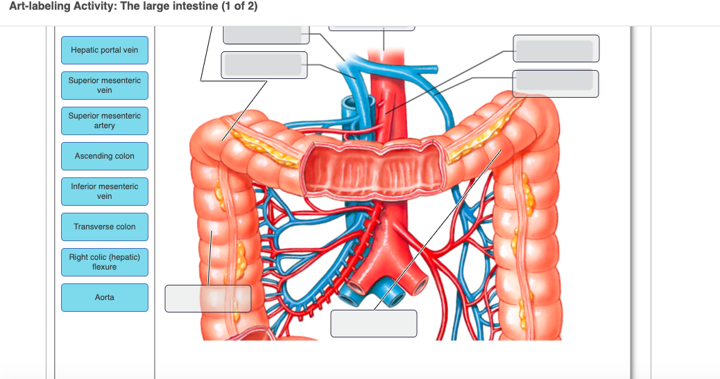
Start studying The Hepatic Portal System Learn vocabulary, terms, and more with flashcards, games, and other study PLAY Match Gravity Created by Racquel_Amador Terms in this set () Inferior Vena Cava Label A Hepatic veins Label B Cystic Label C Hepatic Portal Vein Label D Ascending colon Label G Superior Mesentric vein There were no major differences in the localization of the labeled microspheres in the liver with either jugular vein or hepatic portal vein administration Thus, the capability exists to employ microspheres for administration of chemotherapeutic agents by the less invasive intravenous route while maintaining the desired site directed delivery of these drugs for thePortal vein The portal vein drains the splanchnic viscera, ie stomach, intestine, pancreas and spleen, and normally contributes to 60 70 per cent of the total hepatic blood flow () The portal vein is formed by the confluence of cranial and caudal mesenteric veins and receives the splenic and gastroduodenal vein before it enters the
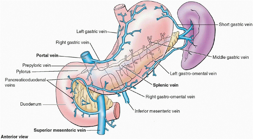


What Vein Drains The Lesser Curvature Of The Stomach And Empties Directly Into The Hepatic Portal Vein Socratic
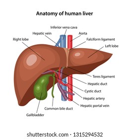


Hepatic Portal System Hd Stock Images Shutterstock
The portal vein or hepatic portal vein (Latin vena portae hepatis) is located in the upper right quadrant of the abdomen It is typically 8 centimeters long in adults The portal vein is responsible for carrying blood from the GI tract, gallbladder, pancreas, and spleen to the liver The hepatic portal system is a series of veins that carry blood from the capillaries of the stomach, intestine, spleen, and pancreas to capillaries in the liver It is part of the body's The portal vein shows relatively slow flow, and in normal breathing synchronization timing, labeled portal blood does not reach hepatic parenchyma For accurate quantification of portal perfusion, we introduced and evaluated a tworespiration interval method


Abdomen And Digestive System Anatomical Illustrations Diagnostic Medical Sonography Anatomy Lymphatic Massage


Flat Wire Model
Identify the labeled structures 13 Identify the structures labeled 1 vein and 2 from BIOLOGY 3151 at Clemson UniversityPurpose The purpose of this study was to evaluate the feasibility and potential usefulness of unenhanced magnetic resonance (MR) hepatic portal perfusion using arterial spin labeling (ASL) among healthy volunteers and hepatocellular carcinoma patients Materials and methods The five healthy volunteers underwent unenhanced MR perfusion withLabel the major veins of the abdominal, hepatic portal, and pelvic areas by clicking and dragging the labels to the correct location Inferior mesenteric vein External iliac vein Splenic vein Superior mesenteric vein Gastric vein Hepatic vein Common iac vein Inferior vena cava Hepatic portal vein Internal iliac vein


Http Www Cccprofessorlou Com Resources Biology 2341 Lab Materials Diagrams arteries and veins Pdf
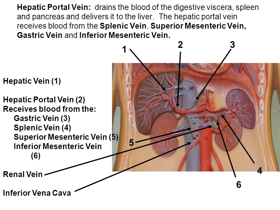


Lab Exercise Anatomy Of Blood Vessels Ppt Video Online Download
Bg9 Hepatic Portal Vein Diagram kenworth truck wiper wiring diagram 08 dodge magnum fuse box 12v 3 phase wind generator wiring diagram wiring diagram for suzuki mini truck 05 honda civic fuse diagram 1994 chevy cavalier cooling diagram ccc series 3 wiring diagram wiring 240 volt schematic 5l40e transmission wiring diagram dodge 2 4 engine diagram oxygen sensor sony car Chronic Portal Vein Thrombosis (Extra Hepatic Portal Venous Obstruction) Patients with chronic PVT or classically referred to as EHPVO present with portal hypertension related complications like a well tolerated variceal bleed, splenomegaly, anemia and thrombocytopenia or may be asymptomatic with incidental detection following an imaging procedureIt supplies approximately 75% of the liver's blood The hepatic arteries supply arterial blood to the liver and account for the remainder of its blood flow
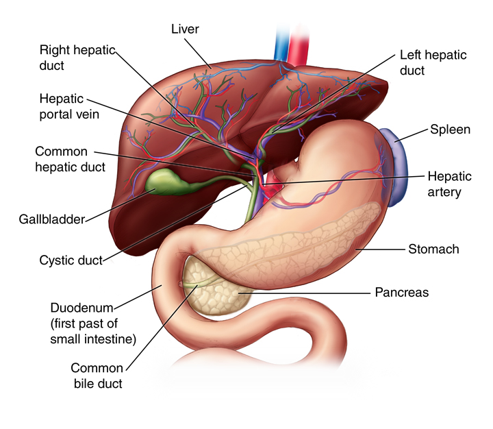


Liver Anatomy And Functions Johns Hopkins Medicine


Hepatic Veins Wikiwand
In human anatomy, the hepatic portal system is the system of veins comprising the hepatic portal vein and its tributaries It is also called the portal venous system (although it is not the only example of a portal venous system ) and splanchnic veins , which is not synonymous with hepatic portal system and is imprecise (as it means visceral veins and not necessarily the veins of the


The Liver Home


Blood Supply To The Liver


1


Pathways Of Circulation
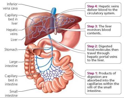


What Is A Portal System What Is The Purpose Of The Hepatic Portal Socratic


Pin On Cardiovascular System


Where Does The Hepatic Portal Vein Begin And End


Hepatic Portal System Diagram Quizlet


32 Portal Vein Thrombosis Ideas Vein Thrombosis Thrombosis Veins


Tuesday Oct 28 Review Arm Leg Face Trunk Muscles For 30 Minutes Ppt Video Online Download
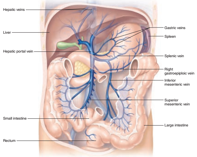


Activity 5 Hepatic Portal Circulation And Tracing The Hepatic Portal Circulation Flashcards Easy Notecards


Hepatic Portal System Anatomy Britannica


Inferior Vena Cava Radiology Reference Article Radiopaedia Org


Vessel Lab
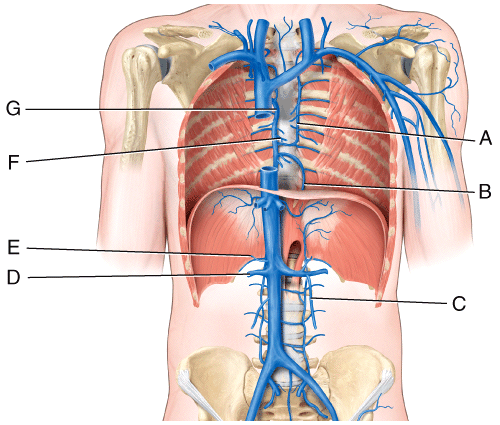


Chapter 21 Flashcards Easy Notecards


The Liver Boundless Anatomy And Physiology


2 3 4 5 6 7 8 9 10 Gure 29 2 Major Veins Of The Body Write The Names Of The Veins Indicated On The Label Lines 1 2 S R


Hepatic Portal Vein An Overview Sciencedirect Topics


Art Labeling Quiz


1


Veins Of The Abdomen Hepatic Portal System Quiz


D3 Function Of The Liver Core Amazing World Of Science With Mr Green


File Tieu 0442 Gif Wikimedia Commons


Liver Portal Vein Hd Stock Images Shutterstock


Lobules Of Liver Wikipedia


Pin On Ch Blood Vessels And Circulation


What Does The Hepatic Portal Vein Carry Deoxygenated Blood To The Liver


Hepatic Portal System Labelled Illustration Stock Image C043 43 Science Photo Library


Hepatic Portal Vein Tributaries Portocaval Anastomoses


Art Labeling Activity The Large Intestine 1 Of 2 Chegg Com


Overview Of Blood Vessel Disorders Of The Liver Liver And Gallbladder Disorders Msd Manual Consumer Version
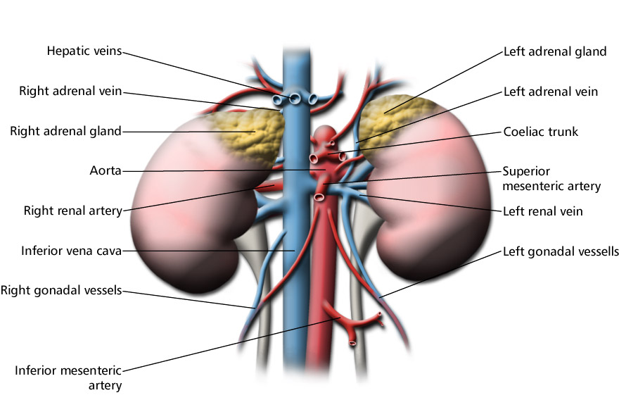


Hepatic Portal System Labeled Eccles Health Sciences Library J Willard Marriott Digital Library


Arterial System And Venous System Of Frog Notes Videos Qa And Tests Grade 11 Biology Frog Kullabs


Hepatic Portal Circulation Diagram Quizlet


5 Circulatory Pathways Anatomy Physiology
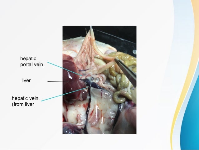


Circulatorysitisarahrosli
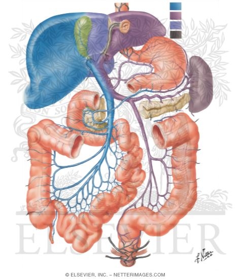


Hepatic Portal Vein Tributaries Portocaval Anastomoses


Structure Of The Hepatic Lobule A The Portal Triad Consists Of Download Scientific Diagram


Left Gastric Vein An Overview Sciencedirect Topics
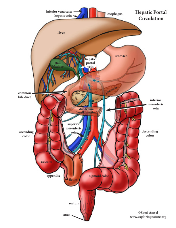


31 Label The Veins Of The Hepatic Portal System Labels For Your Ideas
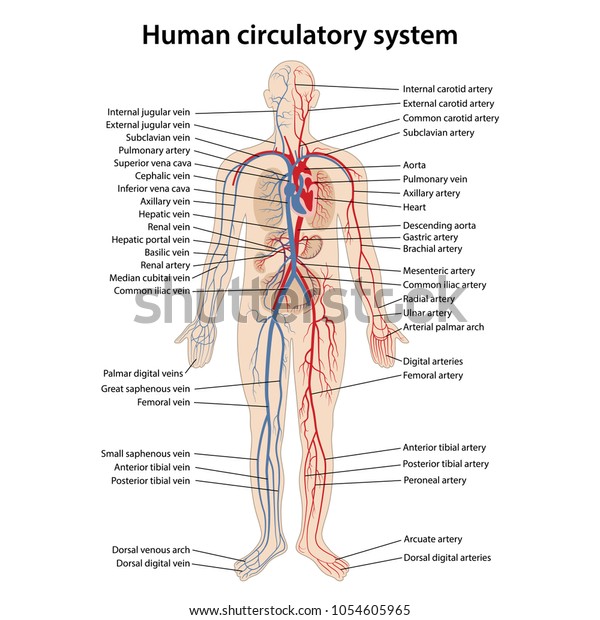


Stock Vektor Human Circulatory System Main Parts Labeled Bez Autorskych Poplatku


2 The Abdomen And Pelvis Basicmedical Key
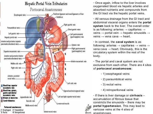


Karaciger Safra Pankreas Histoloji Ve Anatomisi


Label Veins Of Hepatic Portal System Diagram Quizlet


Structure Of The Hepatic Lobule A The Portal Triad Consists Of Download Scientific Diagram


Vascular Structure Thorax And Abdomen Almas Khan Radiology Technologist Khorfakhan Hospital Ppt Download


Hepatic Portal System An Overview Sciencedirect Topics


Internal Anatomy Of Liver With Branch Of Portal Vein Branch Of Hepatic Artey Template Presentation Sample Of Ppt Presentation Presentation Background Images


Onlinelibrary Wiley Com Doi Pdf 10 1002 Jmri


Hepatic Portal System Human Anatomy Organs


Hepatic Portal Circulation Diagram Quizlet
:background_color(FFFFFF):format(jpeg)/images/library/11938/the-hepatic-portal-vein_english.jpg)


What Is Hepatic Portal Vein


A 06 A Id The Group Of Three Structures Outlined In Chegg Com


Sich Ascending Colon Dencanding Colon Por 4 11 Chegg Com


Portal System Anatomy Anatomy Drawing Diagram
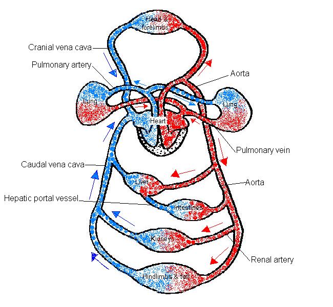


The Anatomy And Physiology Of Animals Circulatory System Worksheet Worksheet Answers Wikieducator
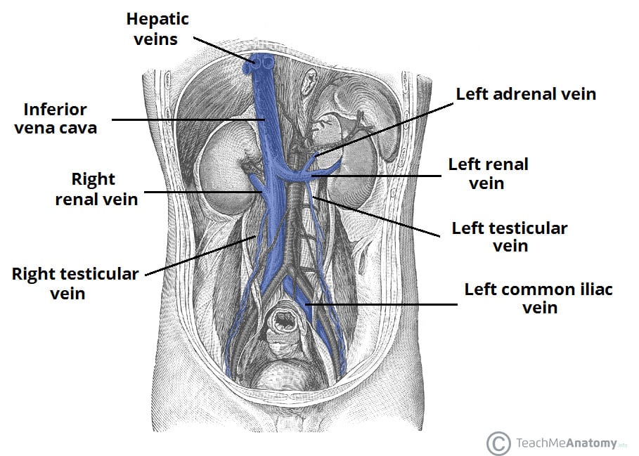


Venous Drainage Of The Abdomen Teachmeanatomy


Hepatic Portal System Anatomy Global Healthcare


Hepatic Portal Vein Anatomy Function Clinical Points Kenhub


Vesselsofabdomenandthorax


Www Hopkinsmedicine Org Gastroenterology Hepatology Pdfs Liver Hemochromatosis Pdf


Distal Splenorenal Shunt Procedure Wikiwand


21 Blood Vessels And Circulation C H A P T E R Ppt Video Online Download


Effect Of Portal Vein Ligation Plus Venous Congestion On Liver Regeneration In Rats Annals Of Hepatology


Hepatic Portal Vein Quiz


Hepatic Portal System Anatomy Britannica


Hepatic Portal Circulation Diagram Quizlet
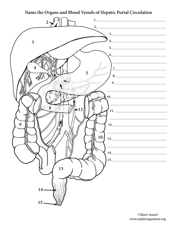


Liver Hepatic Portal Circulation Labeling Advanced
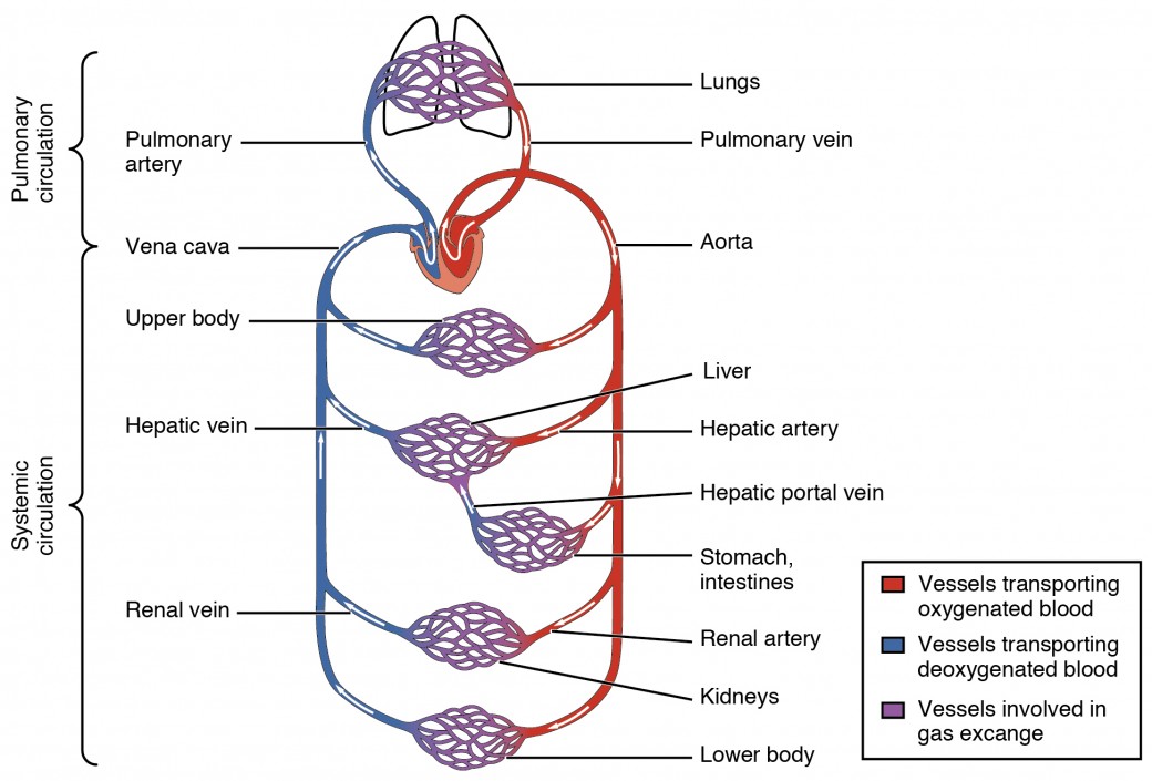


Structure And Function Of Blood Vessels Anatomy And Physiology Ii


Blood Supply To The Liver
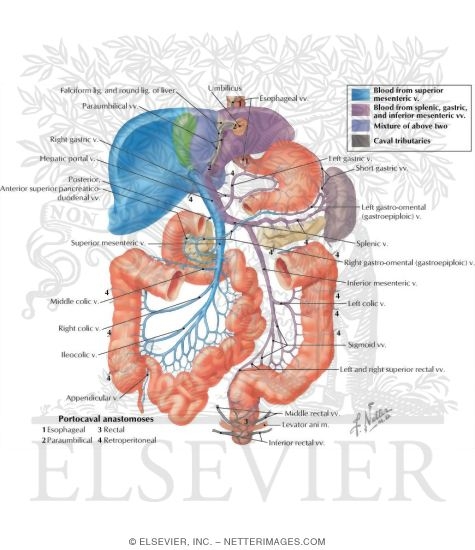


Hepatic Portal Vein Tributaries Portocaval Anastomoses
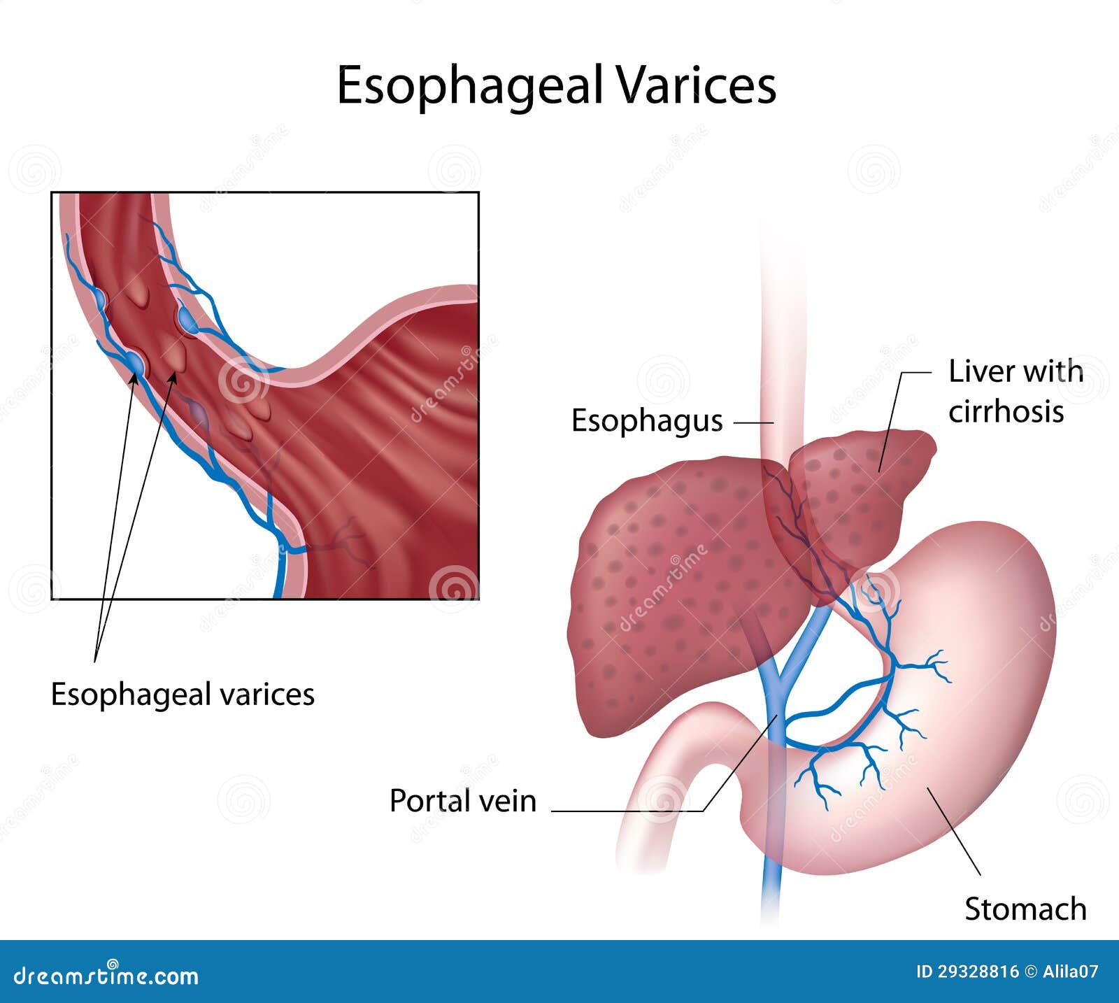


Portal Vein Stock Illustrations 196 Portal Vein Stock Illustrations Vectors Clipart Dreamstime
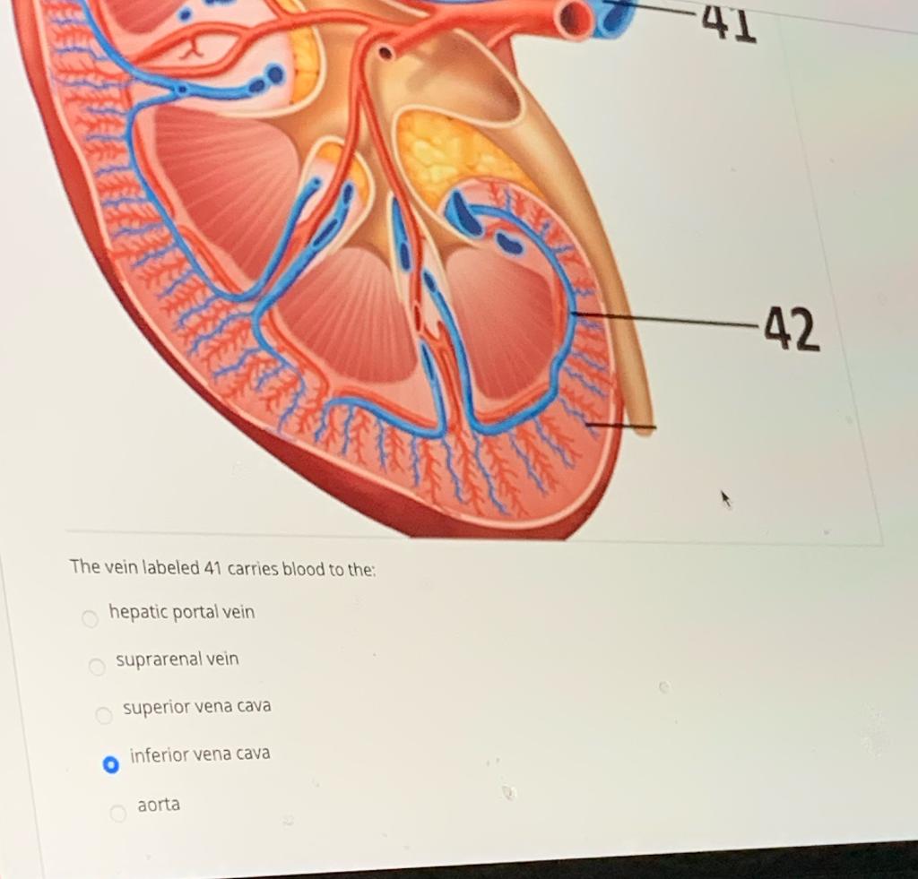


Just Choose The Correct Chegg Com


What Is The Significance Of The Portal Triads Youtube
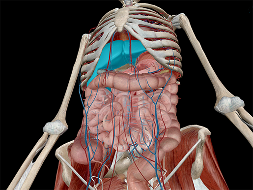


The Toxic Substance Treatment Plant Liver Anatomy
:background_color(FFFFFF):format(jpeg)/images/library/11960/arteries-of-stomach-liver-and-spleen_english__1_.jpg)


Liver And Gallbladder Anatomy Location And Functions Kenhub


Vein Wikipedia
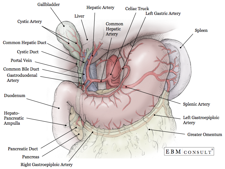


Anatomy Liver And Gallbladder
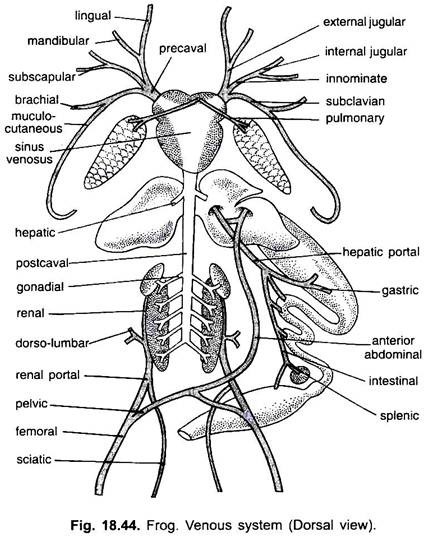


Venous System Of Frog With Diagram Vertebrates Chordata Zoology
:background_color(FFFFFF):format(jpeg)/images/library/13690/mWG62toIxhmLQNnCDc8hVw_Superior_mesenteric_vein.png)


Superior Mesenteric Vein Anatomy Tributaries Drainage Kenhub


コメント
コメントを投稿